The Dyer lab at Emory University studies protein dynamics important in protein folding and enzymatic catalysis with the objective of improving our understanding of human health and disease. We seek to answer fundamental questions such as how atomic motions of the protein structure couple to the catalytic reaction coordinate, and the coupling of backbone and sidechain ordering in a protein folding or misfolding reaction. We are also interested in fundamental problems important to the storage of solar energy as fuel, specifically new photo-catalytic approaches to make hydrogen and to reduce CO2.
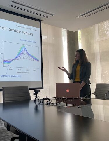
Protein dynamics span a wide range of time scales. We have developed time-resolved spectroscopy methods to investigate protein dynamics from femtoseconds to kiloseconds, with high structural specificity. We emphasize infrared spectroscopy coupled with isotope editing to follow specific biomolecular structural dynamics on all relevant time scales.